Gray matter changes could help explain how childhood trauma triggers PTSD
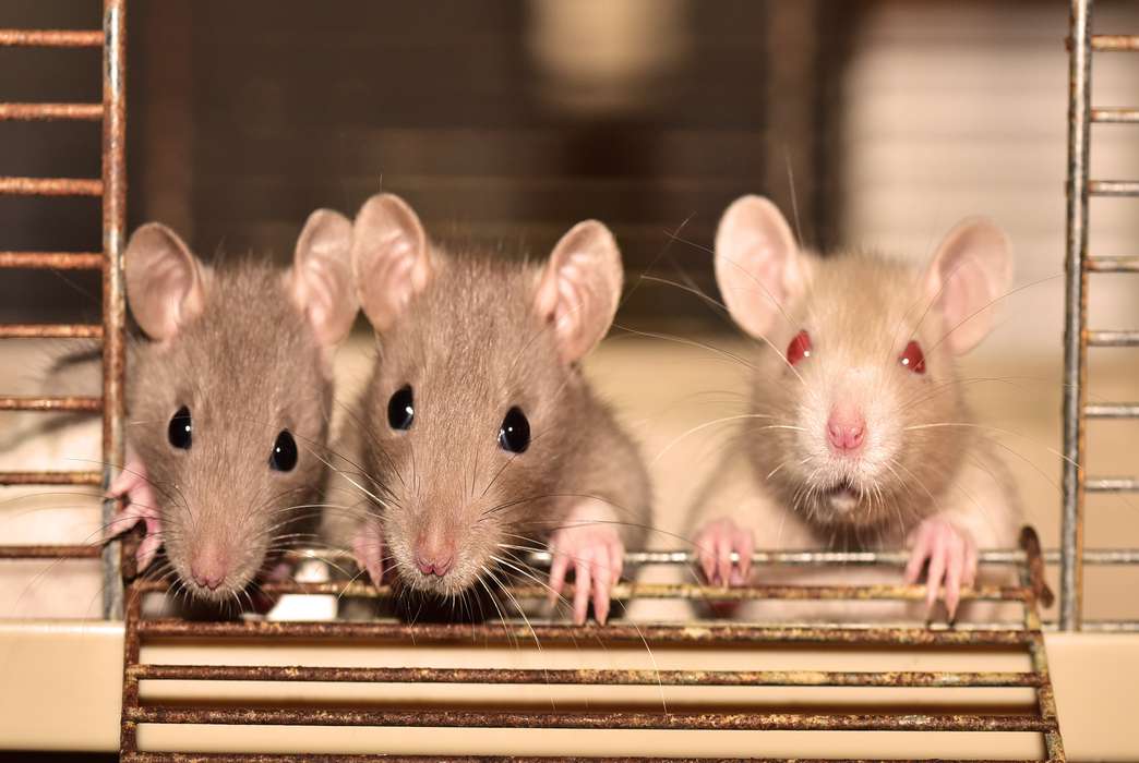
A rat study gives new clues about the damage childhood stress can do that leads to PTSD. (Pixabay/Shutterbug75)

Rats that were exposed to a highly distressing event as juveniles experienced changes in brain regions responsible for processing emotions and detecting danger, offering new clues about the mechanisms that can lead to post-traumatic stress disorder in humans, following adverse events during childhood.
To isolate those brain changes, University of California, Berkeley, researchers tracked the presence of myelin, a mixture of protein and fat that coats nerve fibers, helping to accelerate the transmission of electrical signals in the brain. The team detailed their approach in a Neurobiology of Stress study published April 1.
"A lot of humans will undergo a traumatic experience, but not all will develop PTSD. And that's what we see in our animals, too," Jocelyn M. Breton, formerly affiliated with UC Berkeley, currently a postdoctoral fellow at Columbia University, and the lead author of the study, told The Academic Times. "Similar percentages — around 20% — of our animals will go on to develop this more persistent fear and anxiety-like behavior."
Scientists typically examine myelin levels in the brain's white matter, where myelin accumulates in higher concentrations; white matter is actually named as such because of myelin's white appearance. Gray matter, in contrast, typically contains lower concentrations of myelin, though it is still present to some degree. In fact, this group was the first to study myelin levels in the gray matter of the limbic system, a brain region involved in emotion and in threat detection, among juvenile rats exposed to a brief traumatic event.
Changes in myelin levels have been observed in people with a range of psychiatric disorders, from depression to schizophrenia. The brains of children with ADHD have also been found to display uncommon patterns in white matter. And astronauts who are exposed to microgravity while conducting missions in space may show changes in white and gray matter that are associated with slower recognition of emotions, as well as concentration problems.
Still, the roles of myelin and oligodendrocytes, the cells that help build and regulate myelin, are an underexplored topic area in the field of developmental psychology. Although previous studies have considered how long periods of mild stress alter children's neurological development, few have investigated the impact of a single, highly traumatic scenario, like the kind that can lead to PTSD.
"Using the acute trauma, we're able to pinpoint a little bit further what might be that vulnerable window during development, when the brain is more sensitive to stress," Breton explained. "Maybe it's a region-specific thing. Maybe we would see certain brain regions, like the prefrontal cortex, show sensitivity longer, as it has a longer period of plasticity than other regions."
The UC Berkeley scientists placed juvenile rats in a cone-shaped plastic bag that restricted their movement, leaving them inside the bag for three hours. They also put a cotton ball soaked in fox urine nearby, which led the rats to believe that a predator was present. The threat of a predator combined with an inability to escape caused the rats to feel an overwhelming sense of fear and stress, which the scientists used to simulate the sorts of traumatic events that can lead to long-term neurological changes in humans.
The brains of male rats showed higher levels of myelin shortly after the traumatic event, with no considerable myelin changes in the long term. Females also showed elevated gray matter myelin shortly after exposure but additionally showed a significant reduction in myelin production in each region of the brain in the long term, two months after exposure. The findings could help explain why, among human populations, females report higher levels of anxiety and PTSD than males, Breton said. Alternatively, the higher levels of myelin could be a protective adaptation, helping female rats cope more effectively than their male counterparts.
But these results should be considered with caution, Breton explained, since there is a great deal of variability among individuals, which could lead to different levels of myelin content, regardless of sex differences.
The findings suggest two possible explanations for how stress could alter myelin levels: Traumatic events could first trigger changes in behavior that gradually lead to different levels of myelin production in the brain. In this sense, myelin could serve as a biomarker to help researchers better identify and isolate potential disorders. Alternatively, acute stress might directly lead to changes in myelin that subsequently alter our behavior. "And that would hint that myelin would actually be a therapeutic target," Breton noted.
Although the team did not have the resources to track the rats' behavior as a factor in the study, Breton said the researchers are interested in exploring how anxiety, fear and reduced levels of pleasure could correlate with myelin levels in future investigations.
Scientists aren't sure how the day-to-day processes of the brain are reshaped by traumatic experiences. One theory is that shifting hormone levels during trauma can prompt oligodendrocytes to mature at faster rates, leading to long-term changes in myelin density. Another hypothesis suggests that heightened neuronal activity during a highly stressful event could accelerate myelin production.
The distressing events in rats also resulted in higher levels of the stress hormone corticosterone, a close equivalent of cortisol in humans, as well as weight loss in the days after the traumatic event. Breton thinks these hormonal surges and physiological changes could affect long-term brain function, especially during critical periods of development, when the brain is especially sensitive to changes in its environment.
As the field of neuroscience gains greater insight into brain plasticity, researchers may be able to more effectively pinpoint how myelin production changes over the course of one's life.
"When we think of brain plasticity, we're not just thinking about neurons anymore," Breton said. "We're also thinking about the glia" — the cells that form myelin — "that are changing in response to the environment and stressors in particular. Getting at that will be really interesting."
The study "Juvenile exposure to acute traumatic stress leads to long-lasting alterations in grey matter myelination in adult female but not male rats," published in Neurobiology of Stress on April 1, was authored by Jocelyn M. Breton, Matthew Barraza, Kelsey Y. Hu, Samantha Joy Frias, and Kimberly L.P. Long, University of California, Berkeley; and Daniela Kaufer, University of California, Berkeley and the Canadian Institute for Advanced Research.
Related sections

Life Sciences, Physical Sciences, Social Sciences
Living near oil wells is linked to impaired lung function
Mind & Behavior, Life Sciences
Brain signature for drug addiction discovered in rats

Social Sciences, Mind & Behavior
Latinos with strong U.S. identity are less immigration friendly

Social Sciences, Business & Economics
How network recruiting may help fix workforce gender imbalances
Life Sciences
Collecting dust could help researchers predict COVID-19 outbreaks
By Miles Martin
Life Sciences, Engineering, Physical Sciences
New breath test invokes machine learning to detect lung cancer
By Asher Jones

Physical Sciences
Flooding in Texas will be intensified by sinking land
Life Sciences
Elusive beaked whale species found to be ‘resilient’ to climate change
By Zack Fishman
Engineering
A smaller, more streamlined design can help virtual reality headsets go mainstream
By Beth Newhart
Mind & Behavior
Why do some TED Talks go viral? It's all about the speaker
By Tara DiMaio