New deep-learning model automatically detects COVID-19 pneumonia lesions
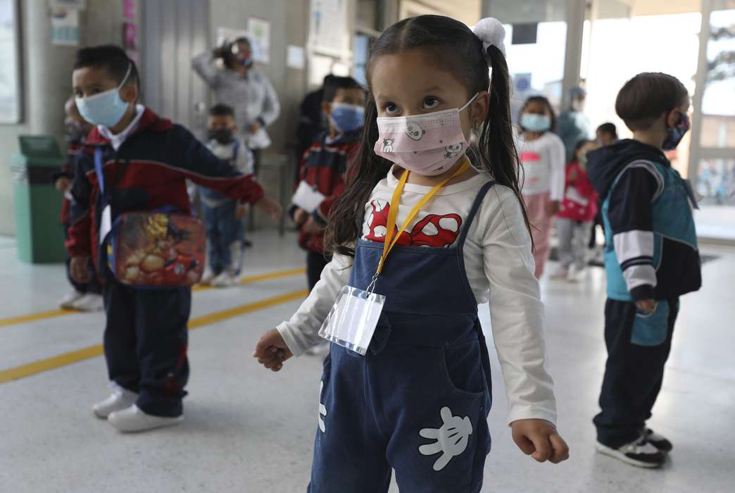
A new technique can detect COVID-19 lung damage with nearly complete accuracy. (AP Image/Sacramento Bee)

Biomedical researchers in Turkey have developed a model for a diagnostic system based on a deep-learning algorithm that can automatically detect lesions in the lungs of patients with COVID-19-related pneumonia through their CT scans with 93.26% accuracy, which can potentially help clinicians diagnose and evaluate pneumonia resulting from the coronavirus.
In a paper published Feb. 16 in the International Journal of Imaging Systems and Technology, the authors explained that they were able to achieve the high level of detection accuracy by successfully segmenting the computed tomography, or CT, images into thousands of smaller images and detecting COVID‐19 lesions in small regions of the lungs.
Previous studies that have used deep learning‐based automatic diagnosis models for lung imaging have only analyzed full or large‐scale regions of the lung image at a time. Mehmet Siraç Özerdem, a professor at Dicle University in Turkey and lead author of the paper, told The Academic Times that the current study is the first to show that lesion patterns can be detected in very small regions of the lung, made possible with their deep learning model.
Deep learning is a subset of machine learning, the study of computer algorithms that are trained how to learn with data and which can improve automatically through experience. Deep learning is inspired by the structure and function of the human brain, and it uses a multilayered structure of algorithms called neural networks, which function much like the human brain in identifying patterns and classifying different types of information.
Most deep-learning models are based on convolutional neural networks, which specialize in analyzing visual imagery. Beyond their use in facial recognition and speech-to-text transcription software, deep learning algorithms have also recently become one of the most popular forms of artificial intelligence at work in the diagnosis of various diseases using biomedical images.
In the current study, the researchers successfully used their deep-learning-based diagnostic model to show that lesion patterns can be detected with a high level of accuracy in small lung regions using CT images. They began this research to help combat some challenges faced by the health care industry caused by COVID-19, as pneumonia is a serious and common complication of the virus.
Detecting lung lesions manually using radiologists is a tiring activity that requires focus and effort, the authors explained, and pneumonia‐induced abnormalities occur in lung CT images in several different forms. The high volume of COVID-19 pneumonia patients and a lack of experienced radiologists can cause diagnostic errors in the interpretation of the images.
“Therefore, the urgent development of automated diagnostic systems that can scan radiological images quickly and accurately is important in combating the pandemic,” the authors said in the paper.
In the study, CT images were taken from two publicly available sources. The researchers used 102 CT scans from 69 people, including from patients who had been diagnosed with COVID‐19 and from healthy patients. A radiologist manually analyzed the CT scans and marked all apparent lesions and abnormalities, which totaled 129.
The CT scans were then broken up into 16,040 smaller image segments. Of these, 10,420 were categorized as healthy lung regions that were COVID-negative, and 5,620 were categorized as regions containing lesions that were COVID-positive.
After the CT image segments were assessed and labeled manually, they were analyzed with the research team’s proposed diagnostic model.
“The developed model managed to detect patterns related to COVID-19 lesions with an accuracy of 93.26%,” Özerdem said. “In addition to this success, the detection of very small sections in the lungs caused by lesions from chest CT images is one of the most important outcomes of the proposed model.”
Özerdem emphasized the importance of creating computer-aided diagnosis systems that can reliably assist doctors with diagnosis and follow-up treatment in COVID-19 patients. Imaging methods such as CT scans and X-rays are the best options for tracking the progress of lung lesions, their changes and any damage caused to the lungs by COVID-19, he said.
Pneumonia cases resulting from the coronavirus are evaluated and treated by radiologists examining abnormalities or lesions in the lungs, but high demand for qualified specialists is stretching the health care sector thin, Özerdem said. The authors said these challenges can be overcome with their novel high-precision, deep learning‐based scanning system.
“The proposed model can be used as an auxiliary system by clinicians in the diagnosis and evaluation of the disease with high accuracy in health centers, where other molecular diagnostic tests are insufficient during the [COVID-19] outbreak,” the authors said.
The study, “Automatic detection and localization of COVID‐19 pneumonia using axial computed tomography images and deep convolutional neural networks,” published Feb. 16 in the International Journal of Imaging Systems and Technology, was authored by Hasan Polat, Bingol University; and Mehmet Siraç Özerdem, Faysal Ekici and Veysi Akpolat, Dicle University.
Related sections
Life Sciences, Engineering
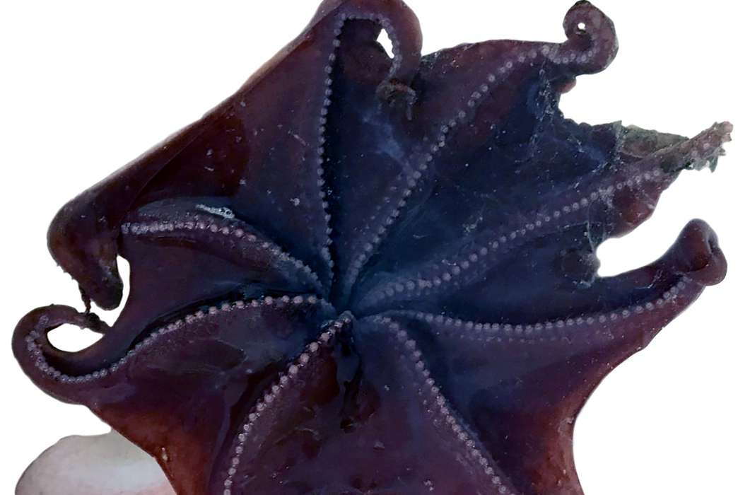
New species of dumbo octopus described through virtual dissections
By Asher Jones

Mind & Behavior, Life Sciences
Scientists find new evidence linking essential oils to seizures
By Beth Newhart
Life Sciences, Mind & Behavior
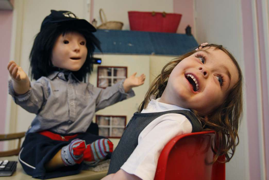
The autistic brain may differ by sex
Physical Sciences, Engineering
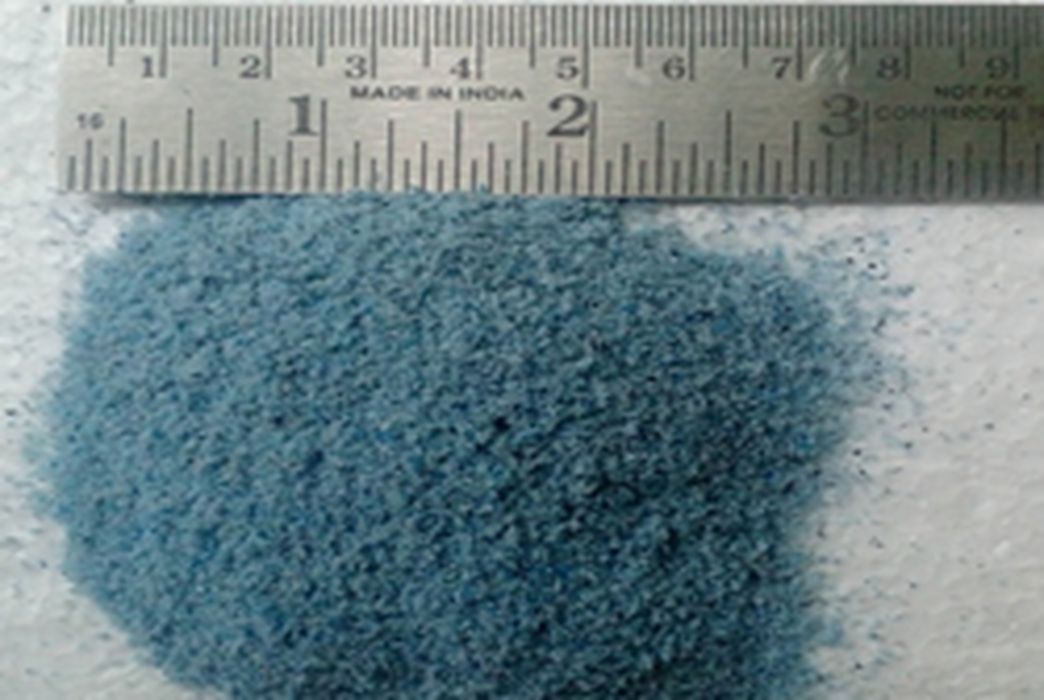
Plastic waste can be put to use in nuclear radiation shielding
By Miles Martin
Business & Economics, Social Sciences, Physical Sciences
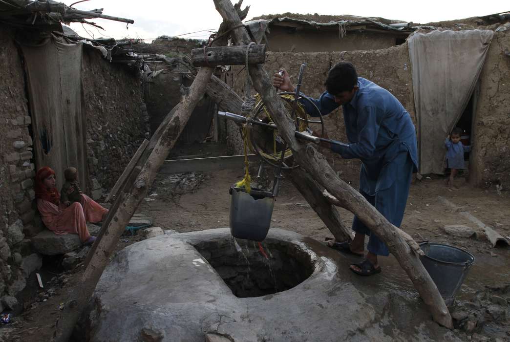
Wells worldwide run risk of running dry, threatening global water supply
Physical Sciences
Alaskan permafrost consistently emits carbon thanks to global warming
By Zack Fishman
Physical Sciences
Astrophysicists found a new way to detect plasma ejections from stars
By Zack Fishman
Mind & Behavior
Kids as young as 4 resemble adults in creative thinking
By Beth Newhart

Life Sciences
This genetic test could help vets prevent untreatable feline kidney disease

Social Sciences, Business & Economics
How local TV can push viewers to the political right
By Theo Wayt